
Radiology and Imaging
The Radiology Department in hospitals uses X-rays, MRIs, CT scans, and ultrasounds to take pictures of the inside of the body. These images help doctors find and diagnose medical conditions early and sometimes assist in treatment.
At Aadhar Hospital, our Radiology & Imaging Department plays a vital role in diagnosing and detecting serious health conditions. Using advanced technology and expert care, our experienced radiologists provide accurate diagnoses to support effective treatment.
We offer a wide range of imaging services, including X-ray, CT scan, MRI, Color Doppler, Mammography, Ultrasonography, and BMD (Bone Mineral Density) scans. With cutting-edge equipment and dedicated specialists, we ensure precise, fast, and reliable imaging for better health outcomes.
Diagnostic Imaging
X- Ray
 X-rays are a fast and effective way to see inside the body, helping doctors diagnose injuries and medical conditions with precision. Using safe radiation technology, X-rays create detailed black-and-white images that our expert radiologists analyze to detect fractures, joint issues, lung infections, and more.
At Aadhar Hospitals, our X-ray services go beyond basic screenings, offering detailed and accurate diagnostics with cutting-edge imaging technology. Every scan is meticulously recorded, ensuring precise and reliable results for your healthcare journey.
What sets us apart is our highly skilled team of radiologists and medical experts, dedicated to delivering clarity and confidence in every diagnosis. With unmatched expertise and advanced technology, we provide seamless, high-quality imaging services to support your well-being.
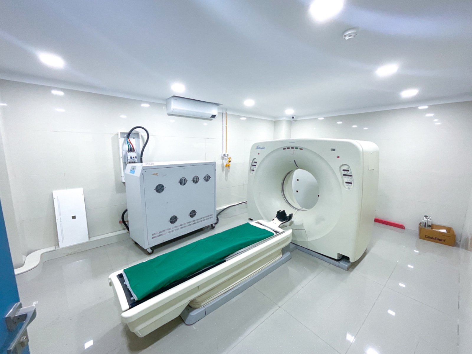
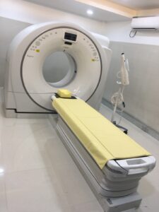
CT Scan
A CT scan is a medical test that helps doctors find diseases and injuries. It uses X-rays and a computer to create detailed images of bones and soft tissues. The scan is painless and does not require surgery. You may have it done at a hospital or an imaging center
The CT Scan Department at Aadhar Hospital is equipped with a state-of-the-art CT scanner, ensuring high-precision imaging for accurate diagnosis. Our skilled and experienced radiologists provide detailed and reliable reports, supporting timely medical decisions. With 24/7 availability, we are committed to delivering efficient and advanced diagnostic services round the clock.
SONOGRAPHY
 Sonography, or ultrasound, is a safe and painless test that uses sound waves to create images of the inside of the body. It helps doctors diagnose and monitor various health conditions without using radiation.
A small handheld device called a transducer sends sound waves into the body. These waves bounce back from different organs and tissues, and a computer turns them into live images on a screen.
- Sonography is commonly used to examine:
- Abdominal organs (liver, kidneys, gallbladder, pancreas)
- The reproductive system (uterus, ovaries, testicles)
- Blood flow in vessels (Doppler ultrasound)
- The heart (echocardiography)
- A baby’s growth during pregnancy
DIGITAL X -RAY AND SONOGRAPHY PROCEDURE
Digital X-ray is a fast, non-invasive imaging technique that uses minimal radiation to capture detailed images of bones and tissues. It helps diagnose fractures, infections, and diseases like cancer.
Sonography procedures include:
- Abdominal Ultrasound: Used to examine organs like the liver, kidneys, and pancreas for conditions such as stones, tumors, or inflammation.
- Pelvic Ultrasound: Primarily used to assess the reproductive organs, including the uterus, ovaries, and bladder in both men and women.
- Obstetric Ultrasound: Commonly used during pregnancy to monitor fetal development, check the health of the baby, and determine the due date.
- Transvaginal Ultrasound: A specialized pelvic ultrasound where a probe is inserted into the vagina to provide more detailed images of the uterus and ovaries.
- Cardiac Ultrasound (Echocardiogram): Used to evaluate heart function, valve issues, and blood flow to detect heart conditions.
- Vascular Ultrasound: Assesses blood flow through arteries and veins, helping diagnose conditions like deep vein thrombosis (DVT) or peripheral artery disease (PAD).
- Musculoskeletal Ultrasound: Helps evaluate muscles, tendons, ligaments, and joints, often used to diagnose injuries or inflammation.
- Thyroid Ultrasound: Used to detect thyroid nodules, cysts, or abnormalities in the thyroid gland.
ADHAR Hospital is equipped with a state-of-the-art ultrasound machine designed for Obstetrics & Gynecological imaging. It features advanced 3D/4D imaging to detect fetal abnormalities, Doppler imaging to assess blood flow, and Fetal Echo for detailed heart evaluations. Additional technologies like I Scan, Panoramic Imaging, and Tissue Harmonic Imaging (THI) enhance clarity, providing precise details of tissue structures and organ vasculature. These advanced tools ensure accurate diagnostics and high-quality care for mothers and babies.